Powered By Blogger

Tuesday, July 31, 2012

HUMAN SKIN

HOME 
SKIN

Skin is outer body covering of an animal. The term skin is commonly used to describe the body covering of any animal but technically refers only to the body covering of vertebrates (animals that have a backbone). The skin has the same basic structure in all vertebrates, including fish, reptiles, birds, and humans and other mammals. This article focuses primarily on human skin.
The skin is essential to a person’s survival. It forms a barrier that helps prevent harmful micro organisms and chemicals from entering the body, and it also prevents the loss of life-sustaining body fluids. It protects the vital structures inside the body from injury and from the potentially damaging ultraviolet rays of the sun. The skin also helps regulate body temperature, excretes some waste products, and is an important sensory organ. It contains various types of specialized nerve cells responsible for the sense of touch.
The skin is the body’s largest organ—that of an average adult male weighs 4.5 to 5 kg (10 to 11 lb) and measures about 2 sq m (22 sq ft) in area. It covers the surface of the body at a thickness of just 1.4 to 4.0 mm (0.06 to 0.16 in). The skin is thickest on areas of the body that regularly rub against objects, such as the palms of the hands and the soles of the feet. Both delicate and resilient, the skin constantly renews itself and has a remarkable ability to repair itself after injury.
STRUCTURE OF SKIN
The skin is made up of two layers, the epidermis and the dermis. The epidermis, the upper or outer layer of the skin, is a tough, waterproof, protective layer. The dermis, or inner layer, is thicker than the epidermis and gives the skin its strength and elasticity. The two layers of the skin are anchored to one another by a thin but complex layer of tissue, known as the basement membrane. This tissue is composed of a series of elaborately interconnecting molecules that act as ropes and grappling hooks to hold the skin together. Below the dermis is the subcutaneous layer, a layer of tissue composed of protein fibers and adipose tissue (fat). Although not part of the skin itself, the subcutaneous layer contains glands and other skin structures, as well as sensory receptors involved in the sense of touch.

HAIR
Hair is a distinguishing characteristic of mammals, a group of vertebrates that includes humans. A thick coat of body hair known as fur protects many mammals from the cold and from the sun’s ultraviolet rays. In humans, a species whose body hair is relatively sparse, this protective function is probably minimal, limited chiefly to the hair on the scalp.


NAILS
Nails on the fingers and toes are made of hard, keratin-filled epidermal cells. They protect the ends of the digits from injury, help us grasp small objects, and enable us to scratch. The part of the nail that is visible is called the nail body, and the portion of the nail body that extends past the end of the digit is called the free edge. Most of the nail body appears pink because of blood flowing in the tissue underneath, but at the base of the body is a pale, semicircular area called the lunula. This area appears white due to an underlying thick layer of epidermis that does not contain blood vessels. The part of the nail that is buried under the skin is called the root. Nails grow as epidermal cells below the nail root and transform into hard nail cells that accumulate at the base of the nail, pushing the rest of the nail forward. Fingernails typically grow 1 mm (0.04 in) per week. Toenails generally grow more slowly.


GLANDS
An adult human has between 1.6 million and 4 million glands, or sweat glands. Most are of a type known as sweat glands, which are found almost all over the surface of the body and are most numerous on the palms and soles. Sweat glands begin deep in the dermis and connect to the surface of the skin by a coiled duct. Cells at the base of the gland secrete sweat, a mixture of water, salt, and small amounts of metabolic waste products. As the sweat moves along the duct, much of the salt is reabsorbed, preventing excessive loss of this vital substance. When sweat reaches the outer surface of the skin, it evaporates, helping to cool the body in hot environments or during physical exertion. In addition, nerve fibers that encircle the sweat glands stimulate the glands in response to fear, excitement, or anxiety. The sweat glands can secrete up to 10 liters (2.6 gallons) of fluid per day, far more than any other type of gland in the body.


Sunday, July 29, 2012

HUMAN EAR

HOME

EAR

Ear is organ of hearing and balance. Only vertebrates, or animals with backbones, have ears. Invertebrate animals, such as jellyfish and insects, lack ears, but have other structures or organs that serve similar functions. The most complex and highly developed ears are those of mammals.

STRUCTURE OF HUMAN EYE

The human ear consists of three sections: the outer ear, the middle ear, and the inner ear. The outer ear includes the auricle (pinna), the visible part of the ear that is attached to the side of the head, and the waxy, dirt-trapping auditory canal. The tympanic membrane (eardrum) separates the external ear from the middle ear, an air-filled cavity. Bridging this cavity are three small bones—the malleus (hammer), the incus (anvil), and the stapes (stirrup). The cochlea and semicircular canals make up the inner ear.


A. OUTER EAR
The outer ear is made up of the auricle, or pinna, and the outer auditory canal. The auricle is the curved part of the ear attached to the side of the head by small ligaments and muscles. It consists largely of elastic cartilage, and its shape helps collect sound waves from the air. The earlobe, or lobule, which hangs from the lower part of the auricle, contains mostly fatty tissue.
The outer auditory canal, which measures about 3 cm (about 1.25 in) in length, is a tubular passageway lined with delicate hairs and small glands that produce a wax-like secretion called cerumen. The canal leads from the auricle to a thin taut membrane called the eardrum or tympanic membrane, which is nearly round in shape and about 10 mm (0.4 in) wide. It is the vibration of the eardrum that sends sound waves deeper into the ear, where they can be processed by complex organs and prepared for transmission to the brain. The cerumen in the outer auditory canal traps and retains dust and dirt that might otherwise end up on the eardrum, impairing its ability to vibrate.
The inner two-thirds of the outer auditory canal is housed by the temporal bone, which also surrounds the middle and inner ear. The temporal bone protects these fragile areas of the ear.
B. MIDDLE EAR

The eardrum separates the outer ear from the middle ear. A narrow passageway called the eustachian tube connects the middle ear to the throat and the back of the nose. The eustachian tube helps keep the eardrum intact by equalizing the pressure between the middle and outer ear. For example, if a person travels from sea level to a mountaintop, where air pressure is lower, the eardrums may cause pain because the air pressure in the middle ear becomes greater than the air pressure in the outer ear. When the person yawns or swallows, the eustachian tube opens, and some of the air in the middle ear passes into the throat, adjusting the pressure in the middle ear to match the pressure in the outer ear. This equalizing of pressure on both sides of the eardrum prevents it from rupturing.
C. INNER EAR
The chain of bones in the middle ear leads into the convoluted structures of the inner ear, or labyrinth, which contains organs of both hearing and balance. The three main structures of the inner ear are the cochlea, the vestibule, and the three semicircular canals.
The cochlea is a coiled tube that bears a close resemblance to the shell of a snail, which is what the word means in Greek. Along its length the cochlea is divided into three fluid-filled canals: the vestibular canal, the cochlear canal, and the tympanic canal. The partition between the cochlear canal and the tympanic canal is called the basilar membrane. Embedded in the basilar membrane is the spiral-shaped organ of Corti. The sensory cells in the organ of Corti have thousands of hairlike projections that receive sound vibrations from the middle ear and send them on to the brain via the auditory nerve. In the brain they are recognized and interpreted as specific sounds.
For example, when the head is upright, the gelatin and mineral particles press down on all the hairlike cells equally. When the head is tilted straight forward by dropping the chin, the gelatin and mineral particles pull on all the hairlike cells equally. If the head is tilted to one side or the other, the cells receive unequal stimulation, varying with the direction and amount of tilt. If the utriculus of both ears is destroyed by injury or disease, the head will hang down limply unless its position can be judged with the eyes. The utriculus is also used to detect the body’s starting or stopping. If a person stops suddenly, the gelatin and mineral particles continue to move, exerting a forward pull on the hairlike cells. The cells then send a specific pattern of nerve impulses to the brain. 

Saturday, July 28, 2012

THE HUMAN EYE

HOME

THE HUMAN EYE

The entire eye, often called the eyeball, is a spherical structure approximately 2.5 cm (about 1 in) in diameter with a pronounced bulge on its forward surface. The outer part of the eye is composed of three layers of tissue. The outside layer is the sclera, a protective coating. It covers about five-sixths of the surface of the eye. At the front of the eyeball, it is continuous with the bulging, transparent cornea. The middle layer of the coating of the eye is the choroid, a vascular layer lining the posterior three-fifths of the eyeball. The choroid is continuous with the ciliary body and with the iris, which lies at the front of the eye. The innermost layer is the light-sensitive retina.
The cornea is a tough, five-layered membrane through which light is admitted to the interior of the eye. Behind the cornea is a chamber filled with clear, watery fluid, the aqueous humor, which separates the cornea from the crystalline lens. The lens itself is a flattened sphere constructed of a large number of transparent fibers arranged in layers. It is connected by ligaments to a ringlike muscle, called the ciliary muscle, which surrounds it. The ciliary muscle and its surrounding tissues form the ciliary body. This muscle, by flattening the lens or making it more nearly spherical, changes its focal length.

THE FUNCTION OF EYE
Focusing the eye, as mentioned above, is accomplished by a flattening or thickening (rounding) of the lens. The process is known as accommodation. In the normal eye accommodation is not necessary for seeing distant objects. The lens, when flattened by the suspensory ligament, brings such objects to focus on the retina. For nearer objects the lens is increasingly rounded by ciliary muscle contraction, which relaxes the suspensory ligament. A young child can see clearly at a distance as close as 6.3 cm (2.5 in), but with increasing age the lens gradually hardens, so that the limits of close seeing are approximately 15 cm (about 6 in) at the age of 30 and 40 cm (16 in) at the age of 50. In the later years of life most people lose the ability to accommodate their eyes to distances within reading or close working range. This condition, known as presbyopia, can be corrected by the use of special convex lenses for the near range.

 EYE DISEASE

hordeolum
hordeolum
Ptosis
The most common disease of the eyelids is hordeolum, known commonly as a sty, which is an infection of the follicles of the eyelashes, usually caused by infection by staphylococci. Internal sties that occur inside the eyelid and not on its edge are similar infections of the lubricating Meibomian glands. Abscesses of the eyelids are sometimes the result of penetrating wounds. Several congenital defects of the eyelids occasionally occur, including coloboma, or cleft eyelid, and ptosis, a drooping of the upper lid. Among acquired defects are symblepharon, an adhesion of the inner surface of the eyelid to the eyeball, which is most frequently the result of burns. Entropion, the turning of the eyelid inward toward the cornea, and ectropion, the turning of the eyelid outward, can be caused by scars or by spasmodic muscular contractions resulting from chronic irritation. The eyelids also are subject to several diseases of the skin such as eczema and acne, and to both benign and malignant tumors. Another eye disease is infection of the conjunctiva, the mucous membranes covering the inside of the eyelids and the outside of the eyeball.


Respiratory System

HOME

Respiratory System
Respiratory System, in anatomy and physiology, organs that deliver oxygen to the circulatory system for transport to all body cells. Oxygen is essential for cells, which use this vital substance to liberate the energy needed for cellular activities. In addition to supplying oxygen, the respiratory system aids in removing of carbon dioxide, preventing the lethal buildup of this waste product in body tissues. Day-in and day-out, without the prompt of conscious thought, the respiratory system carries out its life-sustaining activities. If the respiratory system’s tasks are interrupted for more than a few minutes, serious, irreversible damage to tissues occurs, followed by the failure of all body systems, and ultimately, death.
While the intake of oxygen and removal of carbon dioxide are the primary functions of the respiratory system, it plays other important roles in the body. The respiratory system helps regulate the balance of acid and base in tissues, a process crucial for the normal functioning of cells. It protects the body against disease-causing organisms and toxic substances inhaled with air. The respiratory system also houses the cells that detect smell, and assists in the production of sounds for speech.

STRUCTURE

The organs of the respiratory system extend from the nose to the lungs and are divided into the upper and lower respiratory tracts. The upper respiratory tract consists of the nose and the pharynx, or throat. The lower respiratory tract includes the larynx, or voice box; the trachea, or windpipe, which splits into two main branches called bronchi; tiny branches of the bronchi called bronchioles; and the lungs, a pair of saclike, spongy organs. The nose, pharynx, larynx, trachea, bronchi, and bronchioles conduct air to and from the lungs. The lungs interact with the circulatory system to deliver oxygen and remove carbon dioxide.
Nasal Passage

The flow of air from outside of the body to the lungs begins with the nose, which is divided into the left and right nasal passages. The nasal passages are lined with a membrane composed primarily of one layer of flat, closely packed cells called epithelial cells. Each epithelial cell is densely fringed with thousands of microscopic cilia, fingerlike extensions of the cells. Interspersed among the epithelial cells are goblet cells, specialized cells that produce mucus, a sticky, thick, moist fluid that coats the epithelial cells and the cilia. Numerous tiny blood vessels called capillaries lie just under the mucous membrane, near the surface of the nasal passages. While transporting air to the pharynx, the nasal passages play two critical roles: they filter the air to remove potentially disease-causing particles; and they moisten and warm the air to protect the structures in the respiratory system. 


Pharynx

Air leaves the nasal passages and flows to the pharynx, a short, funnel-shaped tube about 13 cm (5 in) long that transports air to the larynx. Like the nasal passages, the pharynx is lined with a protective mucous membrane and ciliated cells that remove impurities from the air. In addition to serving as an air passage, the pharynx houses the tonsils, lymphatic tissues that contain white blood cells. The white blood cells attack any disease-causing organisms that escape the hairs, cilia, and mucus of the nasal passages and pharynx. The tonsils are strategically located to prevent these organisms from moving further into the body. One tonsil, called the adenoids, is found high in the rear wall of the pharynx. A pair of tonsils, the palatine tonsils, is located at the back of the pharynx on either side of the tongue. Another pair, the lingual tonsils, is found deep in the pharynx at the base of the tongue. In their battles with disease-causing organisms, the tonsils sometimes become swollen with infection. When the adenoids are swollen, they block the flow of air from the nasal passages to the pharynx, and a person must breathe through the mouth.
Air moves from the pharynx to the larynx, a structure about 5 cm (2 in) long located approximately in the middle of the neck. Several layers of cartilage, a tough and flexible tissue, comprise most of the larynx. A protrusion in the cartilage called the Adam’s apple sometimes enlarges in males during puberty, creating a prominent bulge visible on the neck.
While the primary role of the larynx is to transport air to the trachea, it also serves other functions. It plays a primary role in producing sound; it prevents food and fluid from entering the air passage to cause choking; and its mucous membranes and cilia-bearing cells help filter air. The cilia in the larynx waft airborne particles up toward the pharynx to be swallowed.
Food and fluids from the pharynx usually are prevented from entering the larynx by the epiglottis, a thin, leaflike tissue. The “stem” of the leaf attaches to the front and top of the larynx. When a person is breathing, the epiglottis is held in a vertical position, like an open trap door. When a person swallows, however, a reflex causes the larynx and the epiglottis to move toward each other, forming a protective seal, and food and fluids are routed to the esophagus. If a person is eating or drinking too rapidly, or laughs while swallowing, the swallowing reflex may not work, and food or fluid can enter the larynx. Food, fluid, or other substances in the larynx initiate a cough reflex as the body attempts to clear the larynx of the obstruction. If the cough reflex does not work, a person can choke, a life-threatening situation. The Heimlich maneuver is a technique used to clear a blocked larynx (see First Aid). A surgical procedure called a tracheotomy is used to bypass the larynx and get air to the trachea in extreme cases of choking.


FEMALE REPRODUCTIVE SYSTEM

HOME

FEMALE REPRODUCTIVE SYSTEM

The female reproductive system is more complex than that of the male reproductive system. In addition to producing female sex cells, known as ova (eggs), the female body also protects and nurtures a developing baby for about nine months while it grows within the woman’s uterus (womb). A woman’s breasts can provide nourishment for newborns.

BREST

During pregnancy, breast size increases as milk-producing glands known as lobules develop in preparation for breast-feeding. Breast milk can provide all the nourishment a newborn needs. As a newborn breast-feeds, the sucking action stimulates the release of oxytocin, a hormone that promotes milk flow.


OVARIES

The primary reproductive organs of a female are the ovaries, a pair of almond- shaped glands. At puberty, ovaries produce about 400,000 eggs. Each month an egg is released from the ovary and travels down one of the fallopian tubes. If the egg is released around the time of sexual intercourse and it meets and fuses with a male’s sperm, it becomes fertilized. If an egg is not fertilized, it moves from the fallopian tube to the uterus and then passes out of the body in the next menstrual cycle.

 
UTERUS

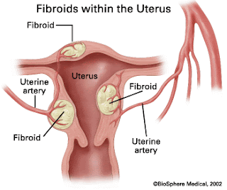
The uterus is a hollow, muscular organ located on top of the bladder. During pregnancy, a fertilized egg travels to the uterus, embeds within the uterine wall, and forms a fetus . Around 40 weeks after fertilization, the fetus is born. During birth, the baby leaves the uterus and travels through the mother’s vagina, a tube that extends from the uterus to the outside of the body.